MEDICAL PHYSICS: The Effects of Radiations and Imaging Using Ultrasound.
Most children playing with an electric torch
have noticed the red glow that comes through their hand when they put it over
the lens. Light passes through quite easily, but it doesn’t give a clear
picture of what is under the skin because the soft tissue scatters light.
An X-ray image of a hand is a fine-tuned version of the picture using a torchlight. Blood vessels can be shown clearly when the blood is injected with a dye that strongly absorbs photons.
A different picture is obtained when a very short pulse of infrared light from a laser is used. The light that penetrates the hand and comes straight out the other side is detected in an equally short time, while scattered photons, which take longer to pass through the hand because they travel further, are not recorded. So the resulting image is sharp.
Images from infrared are not yet as clear as X-ray images because resolution with this technique is still poor. However, they can be useful because the colours indicate the quantity of haemoglobin (the oxygen-carrying compound) in blood. An advantage is that infrared radiation does not ionize atoms, so it is safer to use than X-rays.
With improvements such as computer enhancement giving better resolution (detail) of images, both infrared and visible light imaging could become very safe techniques for both patients and medical workers.
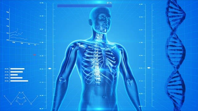
This article deals with the use of physics to diagnose (Part A) and treat (Part B) illnesses and other medical conditions that mar a person’s life, happiness and comfort. Most of the techniques described use radiations of one kind or another, covering most of the electromagnetic spectrum. Even radio is used, to supply energy to the oscillating magnetic fields used in magnetic resonance imaging (MRI). Some techniques involve the use of particles such as beta particles and positrons, emitted by radioactive elements.
To use all these radiations, specialist detecting instruments are needed, as well as highly technical means for producing and directing the beams.
Medical physicists need to be aware, not only of the beneficial effects of radiations but also of the possible harmful effects that they might have on patients and on medical personnel.
PART A: PHYSICS IN MEDICAL DIAGNOSIS
A trained observer can find out a great deal about the state of health of the human body by looking at it, examining by touching it, studying substances from it, and listening to its internal noises. Usually, the patient can also describe the nature and site of any pain. But until the end of the nineteenth century, finding out more required invasive techniques, such as cutting an organ open in an exploratory operation. This carried the risk of trauma (damage and shock) and infection. But in 1895 Wilhelm Konrad Röntgen discovered X-rays. He saw that they cast a shadow of a hand on a luminous screen. One of his X-ray images revealed the bones and the ring on a finger of his wife’s hand. Within weeks of the discovery, doctors were using home-made X-ray machines to look at broken bones and other internal structures. They did so with dangerous enthusiasm, not knowing that X-rays were ionising radiations that could harm living tissue.
X-rays are an example of a non-invasive method for probing deep into the body without cutting into it. Doctors now use a wide range of non-invasive (and often less risky) techniques, both as probes for diagnosis, and also to provide treatment. These involve not only X-rays but also high-frequency sound waves (ultrasound), high-frequency radio waves, light, infrared and ultraviolet radiation and subatomic particles.
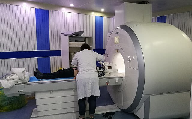
Patient being positioned for MR study of the head and abdomen.
Ptrump16 - Own work, CC BY-SA 4.0
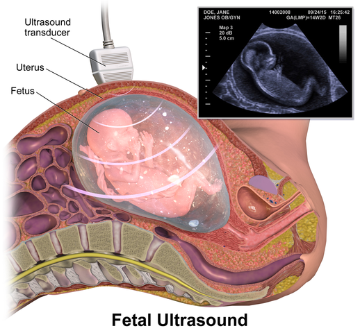
IMAGING USING ULTRASOUND
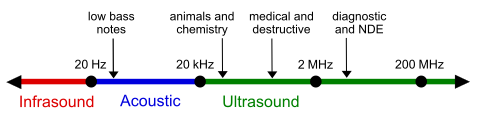
Ultrasound waves are longitudinal pressure waves at a frequency well beyond the upper limit of human hearing (>20 kHz). Low-intensity ultrasonic waves pass through tissue without causing harm and are reflected at the boundaries between different biological structures. These reflections allow images (body scans) of internal organs to be created by an ultrasound scanner.
THE PIEZOELECTRIC TRANSDUCER
Typical diagnostic frequencies are in the range 1 to 5 MHz. Such high frequencies are produced by an electromechanical effect known as piezoelectricity. Certain crystals, such as quartz (SiO2,), produce an electric charge on their surfaces when compressed. The effect is used in the everyday piezoelectric gas igniter: squeezing the handle compresses a crystal and the charge produced makes a spark to ignite the gas.
The effect is used in reverse to produce ultrasound. A high-frequency alternating voltage applied to the crystal surface makes the crystal compress and expand at the same frequency, and the crystal’s vibrations generate the ultrasound waves. Vibrations are best at one of the crystal’s natural resonant frequencies – determined by its size and how it has been cut – and so the applied voltage is tuned to match one of the crystal’s resonant frequencies.
A typical ultrasound emitter uses a piezo-crystal of lead zirconate-lead titanate, PZT. It is a more efficient energy converter than quartz, and so can produce higher power output. The emitter also acts as a receiver: incoming waves alter the crystal’s vibrations which, in turn, generate electrical signals. The piezoelectric element can be thought of as a transducer, a device that changes a signal from one energy form to another. As an emitter, it converts electric potentials to mechanical vibrations, and as a receiver, it converts the energy of mechanical vibrations into varying electric potentials.
The ultrasound is produced in short pulses, typically 10 μs long, with a gap of a few hundred microseconds between pulses. Typically, the beam has a mean power of 0.1 mW.

Bats use ultrasounds to navigate in the darkness.
BUILDING AN ULTRASOUND IMAGE
With the transducer in the ‘receive’ mode, the reflections from different layers in the body return before the next pulse is transmitted. The result is displayed on the screen of a cathode ray oscilloscope as a set of line peaks. Each peak shows the position of a reflecting surface and the height of the peak shows how reflective the surface is. This is called an A-scan.
A B-scan produces an image that is easier to interpret. The probe is scanned across the body in a series of lines, just as a TV screen works. The strength and position of the return signal are stored electronically, then transferred to produce an image on a TV screen. The signal strength now controls the brightness – and even the colour – of a spot on the screen, so building up a two-dimensional image.
Just as electric currents experience resistance in a conductor, ultrasound also meets resistance in travelling through a medium like human tissue. This resistance is called specific acoustic impedance, Z. The value of Z depends on two factors: the density of the medium, ρ, and the speed of sound in the medium, c:
Z = cρ
When the sound moves from one medium to another with a different acoustic impedance, some of the wave is reflected back. (Remember that this happens with light, too, when it moves from one medium to another, even when both are transparent.) This is one reason why ultrasound waves do not enter the body easily – not only are they strongly absorbed in the air if there is a gap between emitter and skin, there will also be strong reflection. So a liquid is used to couple the transmitter with the skin – either a thin film of oil or water-based cellulose jelly. But it is this very reflection property that ultrasound imaging relies on. It can be shown that the fraction α of energy that is reflected when ultrasound moves from a medium 1 to a medium 2 is given by the relationship:
α = energy reflected/energy transmitted = (Z2 – Z1)2/(Z2 + Z1)2
Where the Z values are for the two different media.
BruceBlaus - Own work, CC BY-SA 4.0
ULTRASOUND IMAGE QUALITY
As we have seen, the ultrasound image is formed from a set of reflection pulses from material at different depths in the body section scanned. The sharpness of each reflection depends on the duration of the transmitted pulse: if the pulse is too long, then echoes will overlap. But if the pulse is too short, the energy carried is small and the reflections may be too weak to be detected above the background ‘noise’ of the detection system. Thus, the best pulse length is a balance between the two.
Another problem is multiple reflections: the reflected pulse from a boundary may be bounced back into the patient to be reflected once again. This gives multiple overlapping signals which reduce clarity. Also, since the image of an area of the body is built up over time using a scanning procedure, any movement of the patient or internal organ means that linear images taken at different times do not match, and the compound image will be blurred.
Resolution
The image of an object in the body is made up of reflections from small details. Diffraction will occur if the wavelength used is too large. Then, the image will lack clarity, making resolution poor. As a general rule, ultrasound will just resolve details of the same size as its wavelength. This means that if we need to resolve detail to a level of 1 mm, the ultrasound must have a wavelength equal to or smaller than 1 mm. Diagnostic ultrasound uses frequencies in the range 1 to 15 MHz, resolving details as small as 0.1 mm.
BLOOD FLOW MEASUREMENT
One useful application of ultrasound is the measurement of the rate at which blood flows in blood vessels. The method uses the Doppler principle. The ultrasound is reflected by blood particles (red blood cells) and, as the particles are moving, the reflected waves are shifted in frequency by an amount determined by the speed of the blood.
Ultrasound frequencies of 5 to 10 MHz are used to make images. The beam enters the blood vessel at an angle θ. The frequency shift Δf is measured and related to blood speed v by the formula:
Δf = 2fv cos θ/c
Where c is the speed of the ultrasound.
DANGERS OF ULTRASOUND
Ultrasonic waves carry energy. Some of it is absorbed in tissue and causes heating. Bones are particularly energy absorbent. At some frequencies, small objects in the body could resonate and literally be shaken to pieces. So care is taken to keep a low combination of exposure time and intensity.
LightYear at English Wikipedia, CC BY-SA 3.0
Ultrasound can also cause cavitation, the production of small gas bubbles which absorb energy, expand and may damage surrounding tissue. Damage is unlikely at the frequencies and intensities used for diagnosis, but is useful in some kinds of treatment.
Till next time, I remain my humble self, @emperorhassy.
Thanks for reading.
REFERENCES
https://www.encyclopedia.com/medicine/encyclopedias-almanacs-transcripts-and-maps/medical-physics
https://en.wikipedia.org/wiki/Medical_physics
https://en.wikipedia.org/wiki/X-ray
https://en.wikipedia.org/wiki/Medical_ultrasound
https://radiology.ucsf.edu/patient-care/patient-safety/radiation-safety/benefits
https://www.insideradiology.com.au/radiation-risk/
https://www.ncbi.nlm.nih.gov/pmc/articles/PMC4494460/
https://www.hindawi.com/journals/bmri/2018/7569590/
https://www.fda.gov/radiation-emitting-products/medical-imaging/ultrasound-imaging
This post has been voted on by the SteemSTEM curation team and voting trail. It is elligible for support from @curie and @utopian-io.
If you appreciate the work we are doing, then consider supporting our witness stem.witness. Additional witness support to the curie witness and utopian-io witness would be appreciated as well.
For additional information please join us on the SteemSTEM discord and to get to know the rest of the community!
Thanks for having added @steemstem as a beneficiary to your post. This granted you a stronger support from SteemSTEM.
Thanks for having used the steemstem.io app. You got a stronger support!
Hi @emperorhassy!
Your post was upvoted by Utopian.io in cooperation with @steemstem - supporting knowledge, innovation and technological advancement on the Steem Blockchain.
Contribute to Open Source with utopian.io
Learn how to contribute on our website and join the new open source economy.
Want to chat? Join the Utopian Community on Discord https://discord.gg/h52nFrV
Wow! That was a great read! Now I understand why the technician applies gel before an ultrasound. And I never realized that ultrasound could harm. It makes sense, but I always thought of the procedure as non-invasive. In a way, it is invasive, isn't it?
Thanks for offering such an easy-to-understand explanation of a technology most of us have used many times.
No matter what you do, a wave always transports "something". Therefore, the something can or can't be harmful, depending on the cases :)
Well, it's obvious when you explain it. Of course. It just seemed so innocuous. Now I know :)
:)
Your x-ray image (the second one, with the scanner) remembers me some lab classes I was holding back in the days (I taught nuclear physics to medicine students 15 years ago :)
I am looking forward to read the next episode!
Wow.....15 years ago? What a great history!
And here is the next episode u've been looking forward to read, MEDICAL PHYSICS: Imaging with X-rays.
Thanks for coming to read the post, @lemouth.
Yeaaaahhh... I am older than it seems :D