Analysis of the lens system of a compound optical microscope
1.- Analysis and calculations of variables in the generation of images in a convergent lens.
2.- Calculations of variables in image generation in a diverging lens.
4.- Converging-converging lens arrangement in the optical system of a refracting telescope.
5.- Geometry implemented in the understanding of the algebraic formulation of thin lenses.
6.- Analysis of the arrangement of the optical system of a Newtonian reflecting telescope.
7.- Analysis of the arrangement of the optical system of a Newtonian reflecting telescope.
8.- Analysis of the optical elements of a Sextant.
9.- Analysis of the optical elements of a Porro binoculars.
10.- Analysis of the optical elements of a periscope.
11.- Analysis of the optical elements of an SLR camera.
As you have been able to observe, we have made a tour of important optical instruments such as; the refracting and reflecting telescope, Sextant, Porro binoculars, Periscope, Reflex camera and, on this occasion, I will make a general analysis of the optical elements of a compound microscope, this tool has allowed us to visualize elements imperceptible to the human eye and, this thanks to the implementation of lenses that receive the light that carry these small images to our retinas.
This optical instrument allows us to enlarge the image or get as close as possible in order to analyze any tiny sample of our interest and thus to observe any morphological characteristic of cells and tissues, therefore, for our analysis of the light rays, it is necessary to express that this instrument uses lenses in order to increase the light rays passing through a given sample to be analyzed, generating images and from this point of view we will be performing our overall analysis.
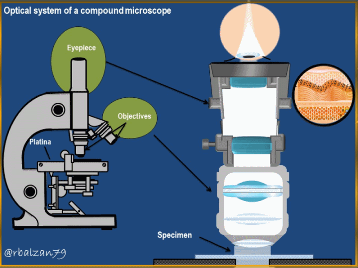
This type of instrument, like the previous optical instruments, has two systems of lenses, some lenses that make up the part of the objective and others that make up the part of the eyepiece, the latter optical system is responsible for bringing the final image to the retina of the person observing through the optical instrument, to begin to relate to these optical systems, it is important that we can see the following figure 1.
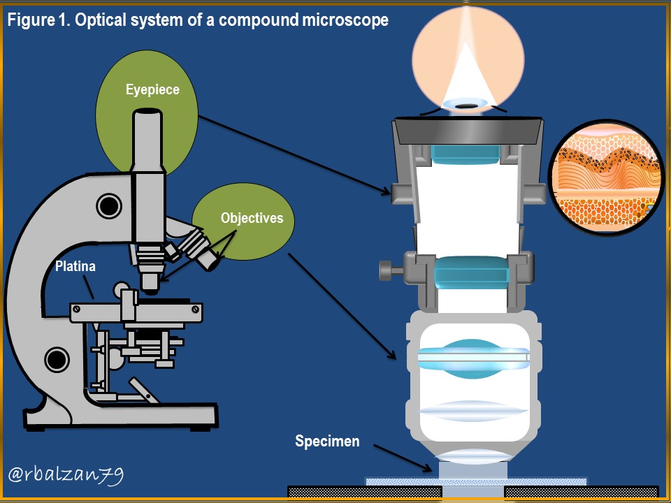
In the previous figure 1, we can observe the optical system of the microscope composed by lenses, both, in its objective part and in its eyepiece part, therefore, let's start to make the schemes of luminous rays that go through each of these convergent lenses, we can observe three lenses in the objective and two lenses in the eyepiece, therefore, let's make the scheme of rays in the lenses present in the objective, therefore, let's observe the following figure 2.
In the previous figure 2, we see the behavior of the images in the three (3) lenses located in the objective part, for the first lens three (3) luminous rays were implemented, the first (red) comes out parallel to the optical axis and, upon impact with the lens is refracted passing through the image focus (F') of the lens, the second ray (black) passes through the center of the lens, so it is not refracted and, a third ray (green), passing through the object focus (F) and upon impact with the lens is refracted parallel to the optical axis, the interception of the three (3) rays generates the image of the object displayed, therefore, we have that the image generated is characteristic of this type of lenses when the observed object is at a distance greater than the focal length, ie inverted image, on the other side of the lens and, real.
For the second lens two light rays were implemented, one of orange color passing through the center of the lens without refracting and the other leaving parallel to the optical axis and, when refracted by the lens passes through the image focus (F'), the intersection of both rays gives us the image, in the same way was done with the third objective lens which is also convergent, that is, using two light rays, the image generated by this third lens was inverted and, with this orientation will reach the first lens of the eyepiece.
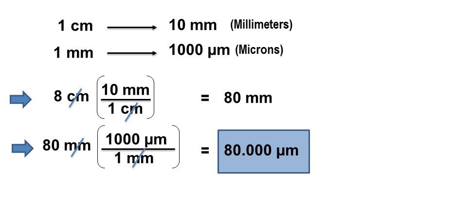
At this point in the path of the luminous rays, that is, through the objective lenses, we will look for or calculate the magnification of the image in the third lens, knowing that the size of the image generated by this lens is 8 cm and the real size of the sample is 2 microns or microns (μm), therefore, to solve the above mentioned, it is necessary to implement the following formula:
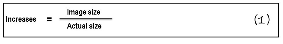
With this formula 1, we look for the data we have, i.e., the following data:
Image size = 8 cm.
Actual size = 2 μm.
In this way, we only need to substitute the values in our formula 1, but first it is necessary to transform or convert the units from cm to μm or vice versa, in this case we will do it from cm to μm, therefore, we proceed first to perform this transformation or conversion:
Now we can apply formula 1, in order to calculate the magnifications experienced by the sample through the three converging lenses of the objective:
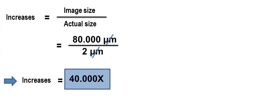
Therefore, we obtain the number of times that the size of the object or sample is increased through the lenses located in the objective part of this optical instrument, in this case, 40,000 times, now let's continue with the scheme of light rays of the lenses located in the ocular part of this optical instrument (compound microscope), as you can see in Figure 3 below.
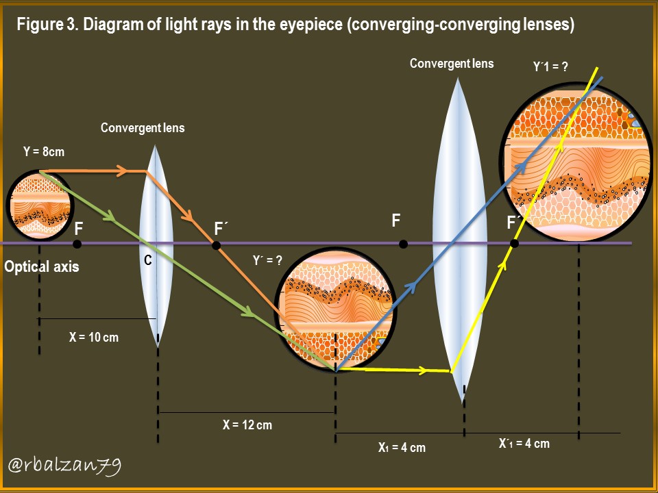
In the previous figure 3, you could observe the ray scheme in the convergent lenses components of the ocular part of the compound microscope, for each lens we use two luminous rays, one that passes through the center of the lens and another ray that leaves parallel to the optical axis and then refract and pass through the image focus (F'), the images are inverted and real, originating on the other side of the lens, i.e. where the light rays that carry the image are transmitted, The images are inverted and real, originating on the other side of the lens, that is, where the light rays that carry the image are transmitted, now we would like to know the size of the final image and for this we have the following data:
Data first convergent lens:
Y = 8 cm (Object height).
X = 10 cm (Object distance).
X´= 12 cm (Image distance).
Y´= ? (Image height).
With these data we proceed to calculate the size of the image generated in the first convergent lens of the eyepiece and, for this purpose, we implement the following formula:
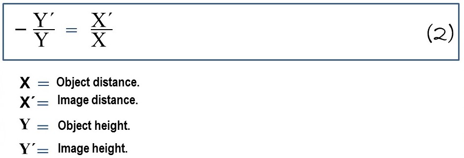
With this formula, we can calculate the size of the image generated by the first convergent lens located in the ocular part of the optical instrument under analysis, therefore, we perform the following procedures:
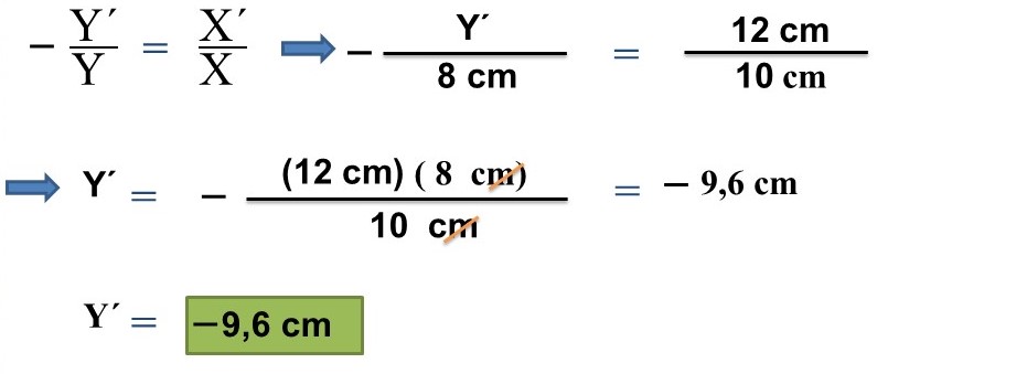
In this way we obtain the image generated by the first convergent lens of the eyepiece, whose value is 9.6 cm, the negative sign indicates that the image is inverted in relation to the orientation of the object, with this value we can continue to the next calculation of the value of the size of the image of the second convergent lens of the eyepiece and, for this, we have the following data:
Second converging lens data:
Y1 = 9,6 cm (Size obtained from the image generated by the first converging lens of the eyepiece).
X1 = 3,2 cm (Object distance).
X´1= 4 cm (Distance of the image generated by the second converging lens of the eyepiece).
Y´1= ? (Height of the final image generated by the second converging lens of the eyepiece).
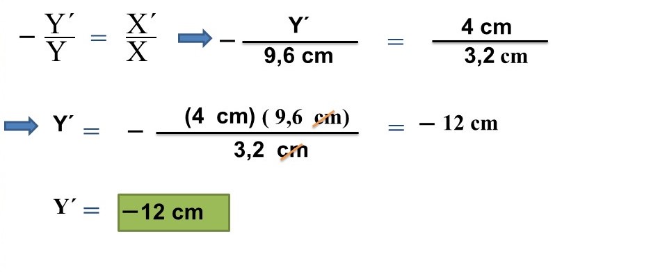
In this way we obtain the size of the final image, that is, the image generated by the second converging lens of the eyepiece of the compound microscope which turned out to be 12 cm, remember that the minus sign (-) means that the image was inverted, characteristic of the converging lenses when the object is at a distance greater than the focal length of the lens, now, we can calculate the final magnification obtained from the sample when passing through each of the converging lenses components of the entire optical system of the compound microscope analyzed.
Using formula 1 and converting from centimeters to microns, we can obtain the proposed value and know how much the lenses were able to increase the size of the sample analyzed, remember that the actual size of the sample is 2 μm and now the size of the final image is 12 cm, with these data we proceed to perform the following calculations:
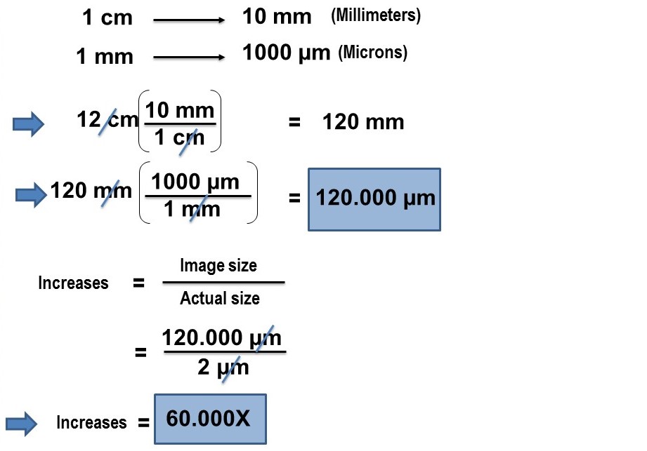
In this way we know the magnification of the final image, that is, the image generated by the second eyepiece lens, whose value was 60,000 x, that is, 60,000 times more than the size of the sample analyzed, remember that the image generated by the third objective lens originated an image of 8 cm, which represented a magnification of 40. Therefore, when the image was passed through the eyepiece lenses, this size increased to 12 cm, representing 20,000 times more, i.e. 50% more than the image generated in the objective lenses.
Conclusion
Undoubtedly, each of the optical instruments designed by man, have fulfilled an essential task in our development as humanity, of course, thanks to the tireless work of the field of science, where, the phenomenon of light, represents one of many phenomena analyzed and interpreted by this field and, This task is still in force and will be so until the end of our days, as we observe how through applied science, that is to say, technology, we are able to take giant steps in the search to increase our longevity on this wonderful planet Earth.
Mathematics and its formulations allow us to learn more about the usefulness of any optical instrument such as the compound microscope, an instrument of great value to capture samples of tissues and cells, which are imperceptible to the human eye, all this thanks to the effect of light and lenses, also thanks to the development of intrinsic phenomena to the propagation of light as is the case of the phenomenon of refraction.
We were able to know the behavior of the luminous rays in 5 convergent lenses, of which three of them, components of the objective part of this optical instrument and, the remaining two of the ocular part, the combination of these five lenses, allowed us to obtain a magnified image of 60,000x, that is, 60 thousand times the size of the real sample, confirming the enormous value of this optical instrument for humanity, for example, in essential areas such as histology and, among other areas of great importance for our evolution.
Until another opportunity my dear friends.
Note: The images were created by the author using Power Point and Paint, the animated gif using PhotoScape.
Recommended bibliographic references
[1] GEOMETRIC OPTICS. Link.
[2] Convergent lenses. Link..
[3] Periscope. Link..
[4] Lens Basics. Link.
[5] Optical microscope. Link.
These are really well analysed
Thank you for your valuable comment. Best regards.
Thanks for your contribution to the STEMsocial community. Feel free to join us on discord to get to know the rest of us!
Please consider delegating to the @stemsocial account (85% of the curation rewards are returned).
You may also include @stemsocial as a beneficiary of the rewards of this post to get a stronger support.
Thank you dear community for your valuable support. Regards.